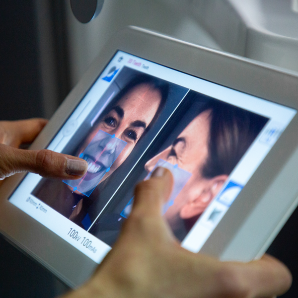
Radiology
Dental radiographies are a standard diagnostic procedure in dentistry, a procedure that provides the dental doctor with significant information about the condition of the teeth and bones for the purpose of preventing and maintaining oral health.


What is dental radiology?
Dental radiology is a branch of dental medicine that is used to diagnose and plan therapies with the help of X-ray images of teeth, jaws and surrounding structures.
It is used for precise analysis of the condition of teeth and bones, assessment of orthodontic irregularities, planning of implant procedures and diagnosis of various pathological changes. Modern digital devices allow minimal radiation exposure with high-quality imaging.
Dental radiology is an excellent diagnostic solution for patients who need an accurate diagnosis of the state of their teeth and jaws, whether it is for routine examinations, planning complex dental procedures, orthodontic therapies, implantology procedures or oral surgery. It is necessary in the most complex dental cases, such as diseases of the temporomandibular joint, sinuses or jaw growth.
FAQ
What is the radiation exposure during CBCT scanning?
CBCT scanning uses a higher dose of radiation than simple dental X-rays (such as bitewing or periapical scans).
However, the radiation dose in CBCT is still significantly lower than that of medical CT (computed tomography) and is relatively low compared to many other medical X-ray methods. The amount of radiation depends on the size of the area to be imaged and the settings of the device.
Are there any risks associated with orthopantomography?
Although there are some risks associated with orthopantomography imaging due to radiation exposure, these risks are minimal when imaging is performed according to guidelines and with adequate protective measures.
Orthopantomography scans are very useful for diagnosis and planning of dental procedures, and their benefits often far outweigh the potential risks.
Can children have orthopantomography?
Children can have orthopantomography, but only when necessary and with adequate protective measures.
How harmful is a dental radiography?
Dental radiography use a very low level of radiation. The level of radiation used in dental x-rays is minimal compared to other sources of ionizing radiation.
To illustrate - a standard dental x-ray is delivered with a dose of radiation equal to the amount of radiation we are exposed to in a day from natural sources such as the Sun or Earth.

"Dental radiology diagnostics are essential for the early diagnosis of dental problems such as cavities, cysts, tumors and abnormalities in tooth development, as well as for the precise planning of procedures such as implants and orthodontic treatments. By using advanced imaging methods, we can be more precise in the diagnosis and adapt the treatments to the specific needs of each patient."
Natalija Gavran, DMD